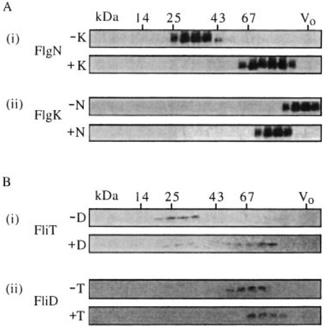
Fig. 4.

In vitro complex formation of FlgN and FliT with substrate HAPs. Gel filtration chromatography of the following purified chaperone and HAP substrate proteins, either alone or after coincubation at a 1:2 molar ratio (substrate–chaperone). Elution fractions from a calibrated Sephacryl S200-HR column were separated by SDS–(15%)PAGE and immunoblotted as indicated. Molecular weight markers are in kDa (see Fig. 2); V0, column void volume.
A. (i) His(6)FlgN alone (−K) and after incubation with His(6)FlgK (+K).Immunoblotted with anti-FlgN antiserum.
(ii) His(6)FlgK alone (−N) and after incubation with His(6)FlgN (+N). [A(ii) bottom shows the same fractions as A(i) bottom]. Immunoblotted with anti-FlgK antiserum.
B. (i) His(6)FliT alone (−D) and after incubation with His(6)FliD (+D). Immunoblotted with anti-FliT antiserum.
(ii) His(6)FliD alone (−T) and after incubation with His(6)FliD (+T). [B(ii) bottom shows the same fractions as B(i) bottom]. Immunoblotted with anti-FliD antiserum.
