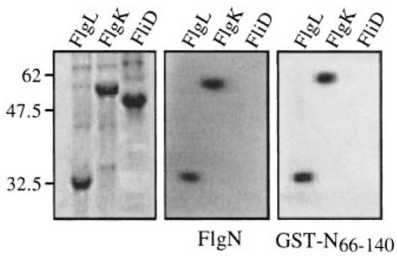
Fig. 6.

Binding of GST-N66–140 to FlgL and FlgK. Late exponential phase cultures of IPTG-induced E. coli BL21 (DE3) carrying the T7 recombinant derivatives pETL (FlgL), pETK (FlgK) or pETD (FliD) were lysed in urea–SDS loading buffer. Proteins were separated by SDS–PAGE (15%) and either stained with Coomassie brilliant blue (left) or transferred onto nitrocellulose membrane and incubated overnight with purified FlgN (middle) or GST-N66–140 (right). Bound proteins were detected by immunoblotting with anti-FlgN polyclonal antisera. Molecular weight markers are in kDa.
