Figure 8.
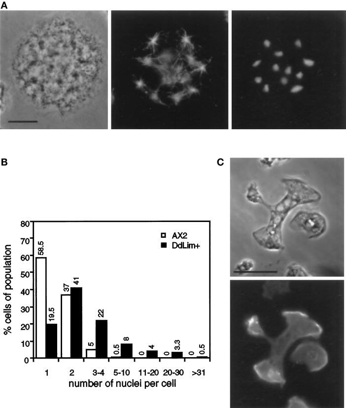
(A) Localization of microtubule and nuclei in a dividing DdLim+ cell. Cells were fixed with cold methanol and stained with anti-α-tubulin (middle) and DAPI (right). Cells were grown in shaking culture for six generations and plated on glass cover slips for 30 min before fixation. Scale bar, 10 μm. (B) Quantitative analysis of the number of nuclei in wild-type Dictyostelium cells (open bars) and DdLim-overproducing cells (solid bars). Cells grown for 12 generations in shaking culture were plated on glass cover slips for 10 min before methanol fixation and nuclei staining with DAPI. The nuclei of 400 cells were counted. (C) Large DdLim-overexpressing cells were allowed to settle for 30 min on a glass coverslip upon which they immediately started to divide by traction-mediated fission. DdLim is located at the leading edge of the separating cell parts.

