Abstract
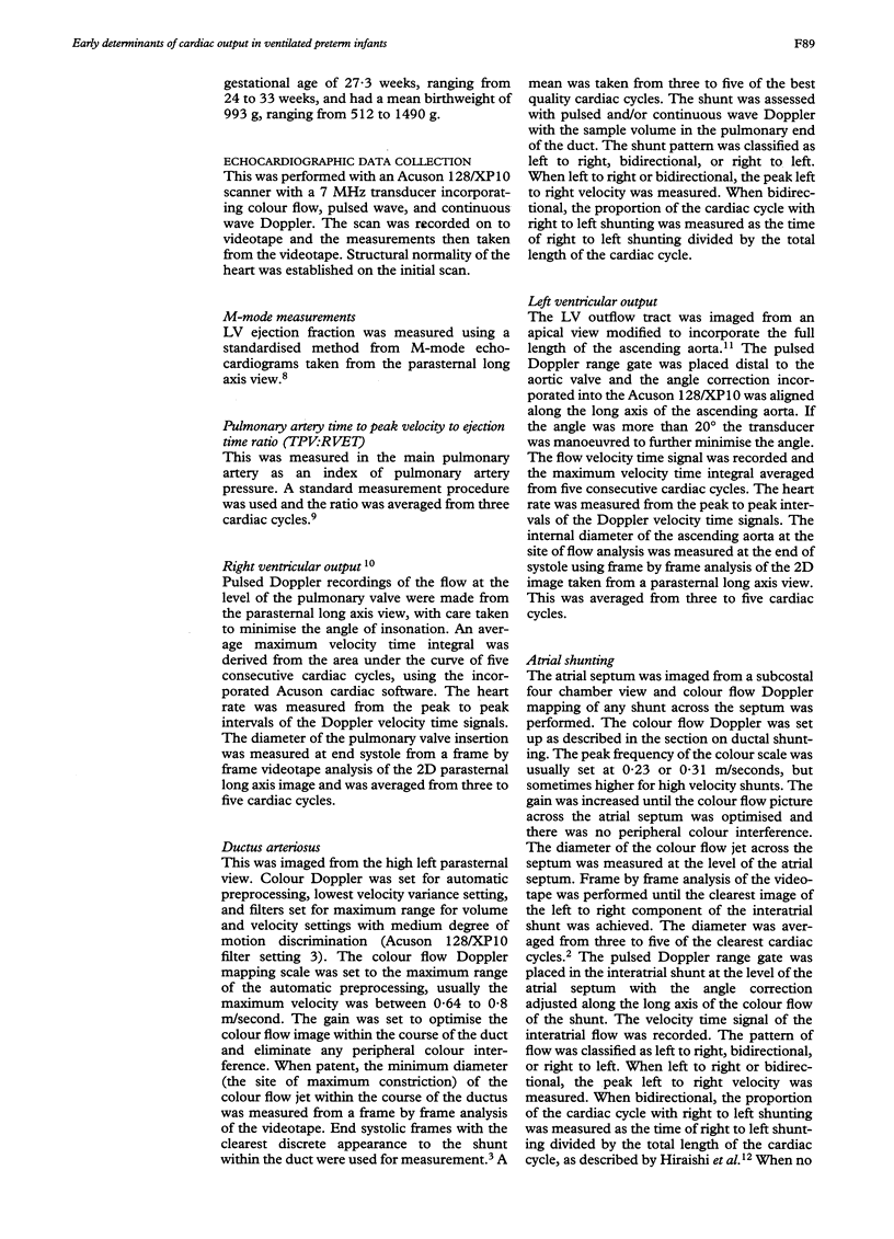
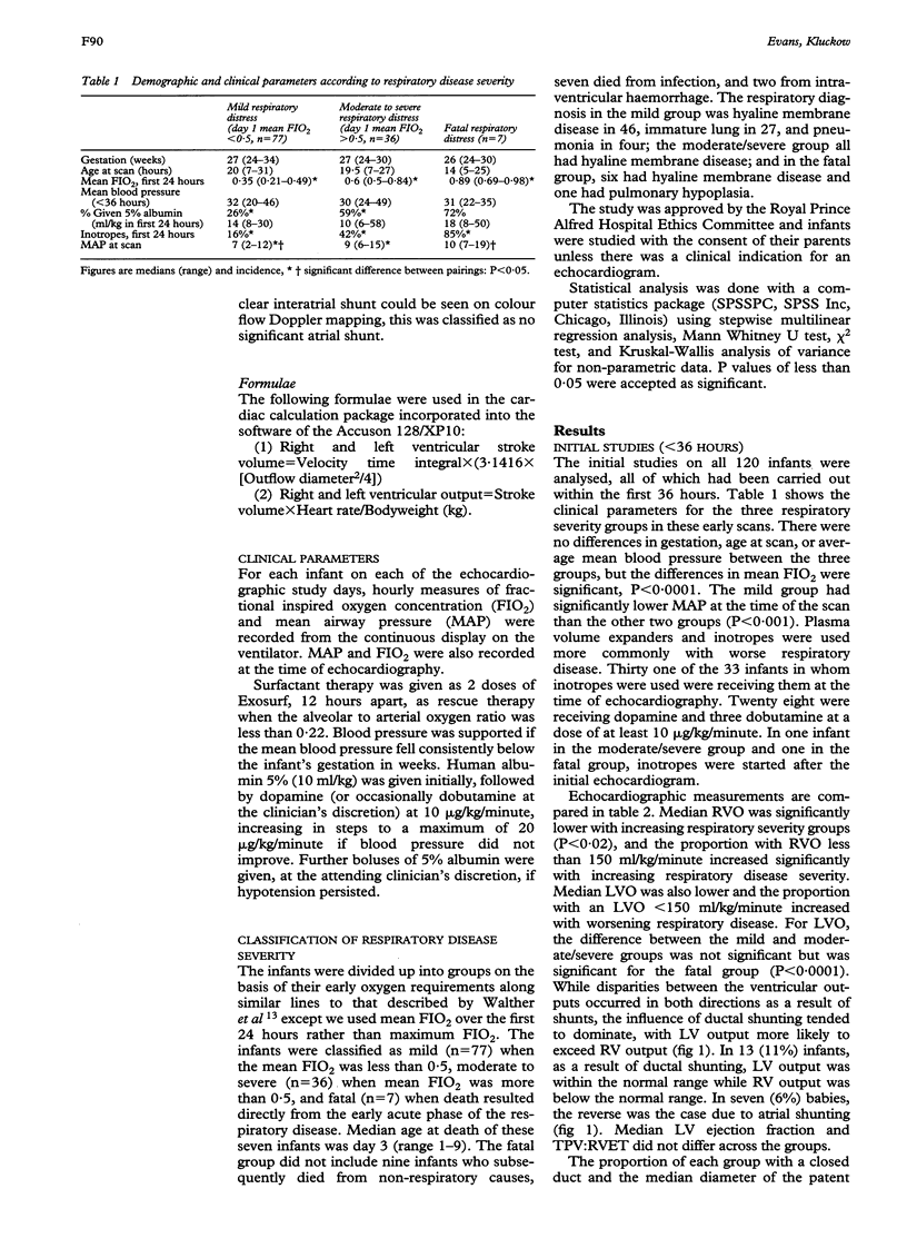
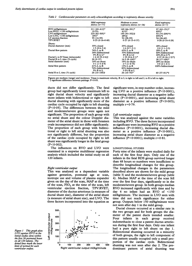
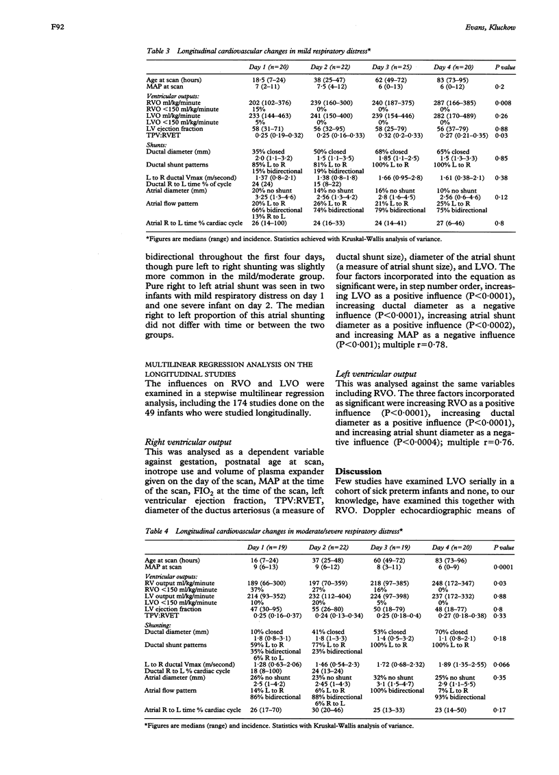
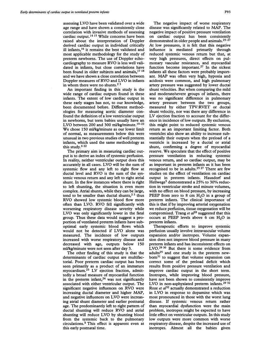
One hundred and twenty ventilated preterm infants, birthweight < 1500 g, were examined within the first 36 hours with colour Doppler echocardiography, to determine the cardiorespiratory influences on right (RVO) and left ventricular output (LVO). Forty nine of these infants had three further daily scans. Measurements included left ventricular (LV) ejection fraction, Doppler determination of RVO and LVO, and ductal and interatrial shunt direction, velocity and colour Doppler diameter. Infants were grouped by respiratory disease severity: mild, mean FIO2 in first 24 hours < 0.5; moderate/severe, mean FIO2 < 0.5; and fatal, death resulting directly from acute respiratory distress. In the early studies ventricular outputs varied widely (RVO: 62-412 ml/kg/minute, LVO: 75-505 ml/kg/minute). The incidence of low ventricular outputs (< 150 ml/kg/minute) increased with worsening respiratory disease. The incidence of low RVO in the mild group was 19%, in the moderate/severe group 42%, and in the fatal group 85%. More infants had a low RVO than a low LVO, reflecting the impact of ductal shunting. Ductal and atrial shunting was predominantly left to right except in those with fatal respiratory disease. In those studied longitudinally, RVO and LVO increased with age and low outputs were not seen after day 3. Multilinear regression analyses, with RVO as the dependent variable, revealed increasing LVO and atrial shunt diameter as significant positive influences and increasing ductal shunt diameter and mean airway pressure as a significant negative influence. With LVO as the dependent variable, increasing RVO, ductal shunt diameter, and age were significant positive influences and increasing atrial shunt diameter was a significant negative influence. Low ventricular outputs are more common with worsening respiratory disease. Mean airway pressure and ductal shunting are two negative influences on ventricular outputs over which there is some therapeutic control.
Full text
PDF
Selected References
These references are in PubMed. This may not be the complete list of references from this article.
- Alverson D. C., Eldridge M. W., Johnson J. D., Aldrich M., Angelus P., Berman W., Jr Noninvasive measurement of cardiac output in healthy preterm and term newborn infants. Am J Perinatol. 1984 Jan;1(2):148–151. doi: 10.1055/s-2007-999991. [DOI] [PubMed] [Google Scholar]
- Alverson D. C., Eldridge M., Dillon T., Yabek S. M., Berman W., Jr Noninvasive pulsed Doppler determination of cardiac output in neonates and children. J Pediatr. 1982 Jul;101(1):46–50. doi: 10.1016/s0022-3476(82)80178-9. [DOI] [PubMed] [Google Scholar]
- Biondi J. W., Schulman D. S., Soufer R., Matthay R. A., Hines R. L., Kay H. R., Barash P. G. The effect of incremental positive end-expiratory pressure on right ventricular hemodynamics and ejection fraction. Anesth Analg. 1988 Feb;67(2):144–151. [PubMed] [Google Scholar]
- Clyman R. I., Mauray F., Heymann M. A., Roman C. Cardiovascular effects of patent ductus arteriosus in preterm lambs with respiratory distress. J Pediatr. 1987 Oct;111(4):579–587. doi: 10.1016/s0022-3476(87)80126-9. [DOI] [PubMed] [Google Scholar]
- Dhainaut J. F., Devaux J. Y., Monsallier J. F., Brunet F., Villemant D., Huyghebaert M. F. Mechanisms of decreased left ventricular preload during continuous positive pressure ventilation in ARDS. Chest. 1986 Jul;90(1):74–80. doi: 10.1378/chest.90.1.74. [DOI] [PubMed] [Google Scholar]
- Evans N. J., Archer L. N. Doppler assessment of pulmonary artery pressure and extrapulmonary shunting in the acute phase of hyaline membrane disease. Arch Dis Child. 1991 Jan;66(1 Spec No):6–11. doi: 10.1136/adc.66.1_spec_no.6. [DOI] [PMC free article] [PubMed] [Google Scholar]
- Evans N., Iyer P. Assessment of ductus arteriosus shunt in preterm infants supported by mechanical ventilation: effect of interatrial shunting. J Pediatr. 1994 Nov;125(5 Pt 1):778–785. doi: 10.1016/s0022-3476(94)70078-8. [DOI] [PubMed] [Google Scholar]
- Evans N., Iyer P. Incompetence of the foramen ovale in preterm infants supported by mechanical ventilation. J Pediatr. 1994 Nov;125(5 Pt 1):786–792. doi: 10.1016/s0022-3476(94)70079-6. [DOI] [PubMed] [Google Scholar]
- Friedman W. F. The intrinsic physiologic properties of the developing heart. Prog Cardiovasc Dis. 1972 Jul-Aug;15(1):87–111. doi: 10.1016/0033-0620(72)90006-0. [DOI] [PubMed] [Google Scholar]
- Gill A. B., Weindling A. M. Echocardiographic assessment of cardiac function in shocked very low birthweight infants. Arch Dis Child. 1993 Jan;68(1 Spec No):17–21. doi: 10.1136/adc.68.1_spec_no.17. [DOI] [PMC free article] [PubMed] [Google Scholar]
- Gill A. B., Weindling A. M. Randomised controlled trial of plasma protein fraction versus dopamine in hypotensive very low birthweight infants. Arch Dis Child. 1993 Sep;69(3 Spec No):284–287. doi: 10.1136/adc.69.3_spec_no.284. [DOI] [PMC free article] [PubMed] [Google Scholar]
- Hausdorf G., Hellwege H. H. Influence of positive end-expiratory pressure on cardiac performance in premature infants: a Doppler-echocardiographic study. Crit Care Med. 1987 Jul;15(7):661–664. doi: 10.1097/00003246-198707000-00007. [DOI] [PubMed] [Google Scholar]
- Hiraishi S., Agata Y., Saito K., Oguchi K., Misawa H., Fujino N., Horiguchi Y., Yashiro K. Interatrial shunt flow profiles in newborn infants: a colour flow and pulsed Doppler echocardiographic study. Br Heart J. 1991 Jan;65(1):41–45. doi: 10.1136/hrt.65.1.41. [DOI] [PMC free article] [PubMed] [Google Scholar]
- Hirsimäki H., Kero P., Wanne O., Erkkola R., Makoi Z. Doppler-derived cardiac output in healthy newborn infants in relation to physiological patency of the ductus arteriosus. Pediatr Cardiol. 1988;9(2):79–83. doi: 10.1007/BF02083704. [DOI] [PubMed] [Google Scholar]
- Lee L. A., Kimball T. R., Daniels S. R., Khoury P., Meyer R. A. Left ventricular mechanics in the preterm infant and their effect on the measurement of cardiac performance. J Pediatr. 1992 Jan;120(1):114–119. doi: 10.1016/s0022-3476(05)80613-4. [DOI] [PubMed] [Google Scholar]
- Maayan C., Eyal F., Mandelberg A., Sapoznikov D., Lewis B. S. Effect of mechanical ventilation and volume loading on left ventricular performance in premature infants with respiratory distress syndrome. Crit Care Med. 1986 Oct;14(10):858–860. doi: 10.1097/00003246-198610000-00004. [DOI] [PubMed] [Google Scholar]
- Mandelbaum-Isken V. H., Linderkamp O. Cardiac output by pulsed Doppler in neonates using the apical window. Pediatr Cardiol. 1991 Jan;12(1):13–16. doi: 10.1007/BF02238491. [DOI] [PubMed] [Google Scholar]
- Mellander M., Sabel K. G., Caidahl K., Solymar L., Eriksson B. Doppler determination of cardiac output in infants and children: comparison with simultaneous thermodilution. Pediatr Cardiol. 1987;8(4):241–246. doi: 10.1007/BF02427536. [DOI] [PubMed] [Google Scholar]
- Notterman D. A., Castello F. V., Steinberg C., Greenwald B. M., O'Loughlin J. E., Gold J. P. A comparison of thermodilution and pulsed Doppler cardiac output measurement in critically ill children. J Pediatr. 1989 Oct;115(4):554–560. doi: 10.1016/s0022-3476(89)80280-x. [DOI] [PubMed] [Google Scholar]
- Rennie J. M. Cerebral blood flow velocity variability after cardiovascular support in premature babies. Arch Dis Child. 1989 Jul;64(7 Spec No):897–901. doi: 10.1136/adc.64.7_spec_no.897. [DOI] [PMC free article] [PubMed] [Google Scholar]
- Rozé J. C., Tohier C., Maingueneau C., Lefèvre M., Mouzard A. Response to dobutamine and dopamine in the hypotensive very preterm infant. Arch Dis Child. 1993 Jul;69(1 Spec No):59–63. doi: 10.1136/adc.69.1_spec_no.59. [DOI] [PMC free article] [PubMed] [Google Scholar]
- Sahn D. J., DeMaria A., Kisslo J., Weyman A. Recommendations regarding quantitation in M-mode echocardiography: results of a survey of echocardiographic measurements. Circulation. 1978 Dec;58(6):1072–1083. doi: 10.1161/01.cir.58.6.1072. [DOI] [PubMed] [Google Scholar]
- Sajkov D., Cowie R. J., Bradley J. A., Mahar L., McEvoy R. D. Validation of new pulsed Doppler echocardiographic techniques for assessment of pulmonary hemodynamics. Chest. 1993 May;103(5):1348–1353. doi: 10.1378/chest.103.5.1348. [DOI] [PubMed] [Google Scholar]
- Sholler G. F., Celermajer J. M., Whight C. M., Bauman A. E. Echo Doppler assessment of cardiac output and its relation to growth in normal infants. Am J Cardiol. 1987 Nov 1;60(13):1112–1116. doi: 10.1016/0002-9149(87)90363-8. [DOI] [PubMed] [Google Scholar]
- Stewart W. J., Jiang L., Mich R., Pandian N., Guerrero J. L., Weyman A. E. Variable effects of changes in flow rate through the aortic, pulmonary and mitral valves on valve area and flow velocity: impact on quantitative Doppler flow calculations. J Am Coll Cardiol. 1985 Sep;6(3):653–662. doi: 10.1016/s0735-1097(85)80127-3. [DOI] [PubMed] [Google Scholar]
- Trang T. T., Tibballs J., Mercier J. C., Beaufils F. Optimization of oxygen transport in mechanically ventilated newborns using oximetry and pulsed Doppler-derived cardiac output. Crit Care Med. 1988 Nov;16(11):1094–1097. doi: 10.1097/00003246-198811000-00002. [DOI] [PubMed] [Google Scholar]
- Walther F. J., Benders M. J., Leighton J. O. Persistent pulmonary hypertension in premature neonates with severe respiratory distress syndrome. Pediatrics. 1992 Dec;90(6):899–904. [PubMed] [Google Scholar]
- Walther F. J., Kim D. H., Ebrahimi M., Siassi B. Pulsed Doppler measurement of left ventricular output as early predictor of symptomatic patent ductus arteriosus in very preterm infants. Biol Neonate. 1989;56(3):121–128. doi: 10.1159/000243112. [DOI] [PubMed] [Google Scholar]
- Walther F. J., Siassi B., Ramadan N. A., Ananda A. K., Wu P. Y. Pulsed Doppler determinations of cardiac output in neonates: normal standards for clinical use. Pediatrics. 1985 Nov;76(5):829–833. [PubMed] [Google Scholar]


