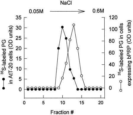
Figure 8.
Ion exchange chromatography of endogenous granule proteoglycan. Medium containing the endogenous granule proteoglycan was collected from [35S]sulfate-labeled cells that had been stimulated with 5 mM 8-Br-cAMP as described in Figure 6. The medium was loaded onto a DEAE cellulose column and eluted with a 0.05 M to 0.6 M NaCl gradient as described in MATERIALS AND METHODS. Equal amounts of each fraction were analyzed by SDS-PAGE and [35S]sulfate-labeled endogenous granule proteoglycan (PG) was quantitated by phosphorimager analysis. Nontransfected AtT-20 cells (•) and cells expressing basic PRP (○).

