
Figure 3.
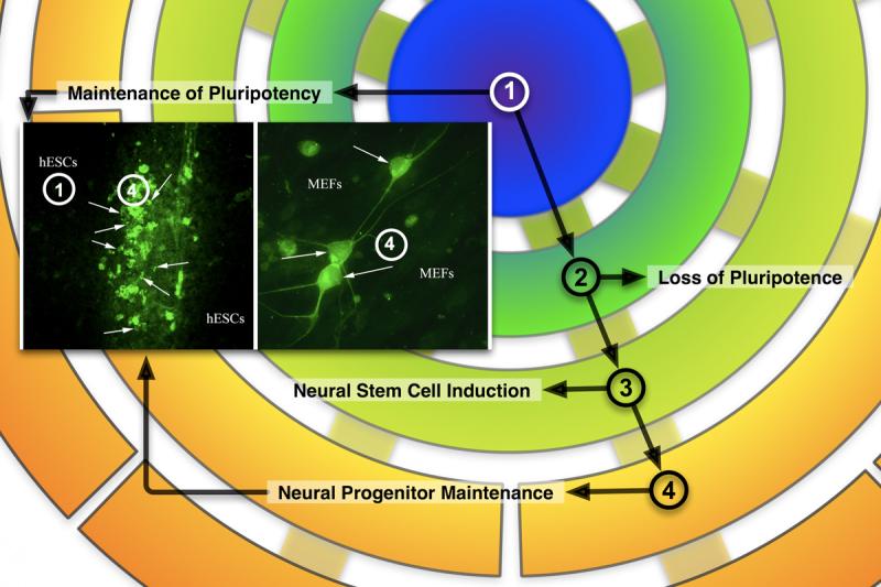
Immunocytochemical staining of doublecortin(DCX)-positive cells (inset) overlaid on the differentiation schema of Figure 1. The inset-left panel (10x) shows DXC+ cells (green, arrows) at the juncture of two colliding PSC colonies (unstained, hESCs), growing under STD conditions. The inset-right panel (40x) shows DCX+ cells (green, arrows) with typical morphologies found in the MEFs (unstained, MEFs) surrounding the colonies. These photomicrographs illustrate that, even under conditions favoring pluripotency, some cells progress through stages 2 and 3 to stage 4 and show, therefore, the propensity of PSCs to differentiate down the neural lineage.