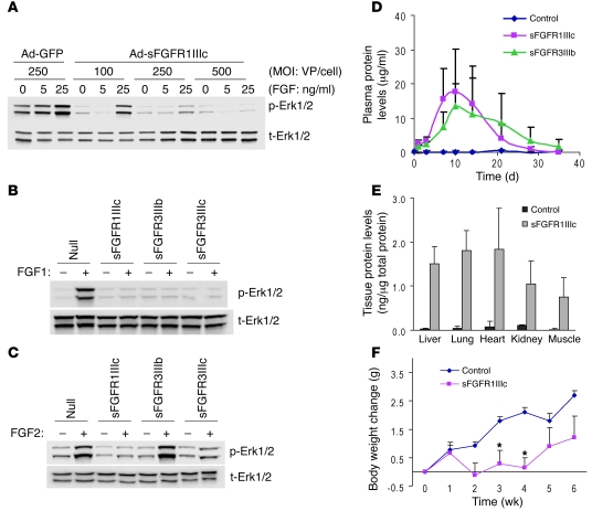
Figure 1. Validation of the sFGFR system.
(A) sFGFR blocks FGF-induced Erk1/2 phosphorylation in vitro in a dose-dependent manner. Ad-GFP and Ad-sFGFR1IIIc were transduced in BAECs and stimulated with indicated concentration of FGF1 for 10 minutes. Total cell lysates were subjected to Western blotting. p-Erk1/2, phospho-Erk1/2; t-Erk1/2, total Erk1/2; VP, viral particles. (B) sFGFRs effectively inhibit FGF1-induced Erk1/2 activation. Ad-Null, Ad-sFGFR1IIIc, Ad-sFGFR3IIIb, and Ad-sFGFR3IIIc were transduced in BAECs and stimulated with FGF1 for 5 minutes. (C) sFGFR3IIIb does not inhibit FGF2-induced Erk1/2 activation. Ad-Null and Ad-sFGFRs were transduced in BAECs and stimulated with FGF2 for 5 minutes. (D) Plasma expression levels of sFGFR in mice. Ad-Null (control) or Ad-sFGFR1IIIc (5 × 10¹⁰ viral particles) was injected into C57BL/6 mice, and blood samples were taken at indicated time points. sFGFR levels were measured using an ELISA system detecting human IgG-Fc. Data are shown as mean ± SD, n = 4 in each group. (E) Tissue distribution of sFGFR. After 10 days of Ad-Null (control) or Ad-sFGFR1IIIc injection (5 × 10¹⁰ viral particles), tissue samples were collected and total protein was extracted. Thereafter, ELISA assays detecting human IgG-Fc were performed. Data are shown as mean ± SD, 4 animals in each group. (F) Body weight change after injection of sFGFR. Ten-week-old C57BL/6 mice were injected with adenoviruses (5 × 10¹⁰ viral particles), and body weight was measured weekly. Data are shown as mean ± SD, n = 4 in each group. *P < 0.05, Student’s t test.

