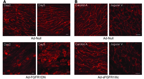
Figure 3. In vivo effect of FGF inhibition in the endothelium.
(A) Enface preparation of the rat femoral artery transduced with Ad-Null or Ad-FGFR1DN (109 PFUs). Segments of arteries were transduced with adenoviruses and stained for VE-cadherin (red) and maximum intensity projection of 1-μm Z-Stack sections is shown. At day 5, endothelial cells lost cell-cell contacts and gaps formed between cells. Scale bars: 10 μm. (B) Mouse carotid artery and jugular vein exposed to systemic Ad-Null or Ad-sFGFR1IIIc viruses. Segments of arteries were stained for VE-cadherin, and maximum intensity projection of 1-μm Z-Stack sections is shown. Scale bars: 20 μm.

