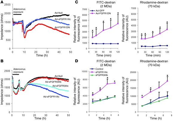
Figure 5. Increased endothelial permeability in cells lacking FGF signaling.
(A) Endothelial monolayer permeability evaluated with ECIS system. Impedance was measured every 5 minutes for 48 hours after the onset of adenoviral transduction. After adenoviral exposure, cell culture medium was changed to normal growth medium. n = 3 in each group, P < 0.05, control versus FGFR1DN or sFGFR1IIIc at 48 hours (B) ESIS analysis using various sFGFR adenoviruses. After adenoviral exposure, medium was replaced with normal growth medium. n = 3 in each group, P < 0.05, control versus sFGFR1IIIc or sFGFR3IIIc at 48 hours (C) Transwell tracer experiment using high (2 MDa) and low (70 kDa) molecular weight dextran. Full confluent BAECs on the Transwell chambers were transduced and dextrans were added in the upper chamber. Fluorescent values in the lower chamber were measured at indicated time points using a fluorescent microplate reader. Three independent experiments were performed. One representative experiment is shown as mean ± SD for n = 3 in each group; *P < 0.05 by Student’s t test, control vs. FGFR1DN. AU represents relative intensity of fluorescence. (D) Transwell tracer experiment using Ad-sFGFRs. Data are shown as mean ± SD for n = 3 in each group; *P < 0.05 by Student’s t test, control vs. sFGFR1IIIc.

