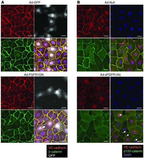
Figure 6. Junction proteins are absent from cell-cell contacts in cells lacking FGF signaling.
(A) Immunostaining of quiescent and fully confluent endothelial monolayer. BAECs were transduced with Ad-GFP or Ad-FGFR1DN-GFP using low MOI (10 and 25, respectively) to limit the number of transduced cells and minimize virus-mediated toxicity. Cells were stained for VE-cadherin (red) and β-catenin (green) 24 hours later. The Ad-FGFR1DN-GFP vector has a bidirectional promoter encoding both GFP and FGFR1DN, thus marking transduced cells with GFP (shown in white). Note the loss of VE-cadherin and β-catenin staining at cell-cell contacts of FGFR1DN-GFP–transduced cells. Arrow points to the gap between neighboring FGFR1DN-GFP–transduced cells. Scale bars: 20 μm. (B) Immunostaining of quiescent endothelial monolayer (BAECs) transduced with Ad-Null or Ad-sFGFR1IIIc. Cells were stained for VE-cadherin (red), p120-catenin (green), and DAPI (blue). sFGFR was secreted in the medium; the effect is not limited to the transduced cells. Arrows indicate gaps between endothelial cells. Scale bars: 20 μm.

