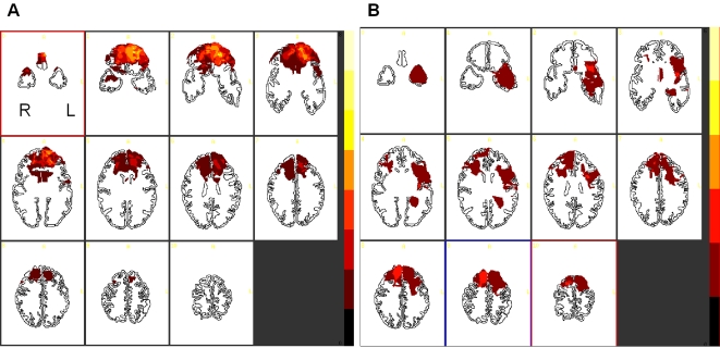
Figure 1. (A). Location and degree of lesion overlap in patients with fronto-polar cortex lesions.
(B). Location and degree of lesion overlap in control patients without fronto-polar cortex lesions. Slices are oriented in radiological convention (i.e. the left side of the image is the right hemisphere). Lighter colors denote the degree to which lesions involve the same structure in multiple subjects. The darker color at the bottom of the color scale indicates no overlap between brain region.

