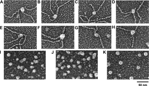
FIGURE 4.
Size analysis of free and DNA-bound WRN proteins. Wild-type (top panel) and K577M (middle panel) WRN were bound to Holliday junctions (A, B, E, and F) and replication forks (C, D, G, and H) and were mounted side-by-side with apoferritin (K) on separate grids for EM analysis along with unbound wild-type (I) and K577M-WRN (J). The projected areas of wild-type and K577M-WRN were measured from EM images and used to estimate the oligomeric forms of free and DNA-bound WRN as described under “Experimental Procedures.”

