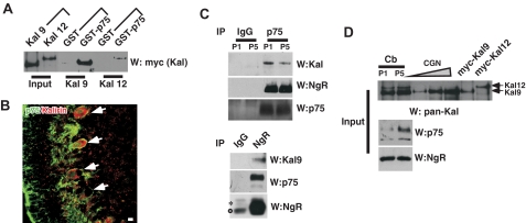
FIGURE 1.
Kalirin9 binds p75. A, the recombinant Kalirin9 and 12 bind the cytoplasmic domain of p75 in vitro. GST or GST-p75ICD was incubated with the lysates from 293T cells that were transfected with the Myc-tagged wild type Kalirin9 or 12. The bound Kalirins were detected by Myc Western analysis (W). For input, 5% of the lysates that were used for GST pulldown were loaded as controls. B, Kalirin is expressed among p75+ Purkinje neurons in the P5 cerebellum. P5 rat sagittal sections were stained with an anti-p75 antibody, 192, and pan-Kalirin antibodies (28). The arrows point to doubly stained Purkinje neurons. Scale bar, 12.5 μm. C, Kalirin9, and not Kalirin12, associates with p75 and NgR in the developing cerebellum. Upper panel, the cerebellar lysates from P5 rat pups were subjected to immunoprecipitation (IP) with a p75 antibody and a control IgG. The bound proteins were simultaneously probed with pan-Kalirin and NgR antibodies using a divided membrane. For detection of p75, the same membrane was stripped and reprobed. *, nonspecific bands. Lower panel, detection of Kalirin9 and p75 in reciprocal immunoprecipitation with NgR antibody. The bound proteins were simultaneously probed with pan-Kalirin and NgR antibodies using a divided membrane. As a control, the same blot was reprobed for NgR following membrane stripping. *, a nonspecific band; ○, IgG heavy chain. D, input controls for C. Note that Kalirin9 is the major isoform present in CGN cultures, whereas Kalirin12 is not expressed at detectable level.

