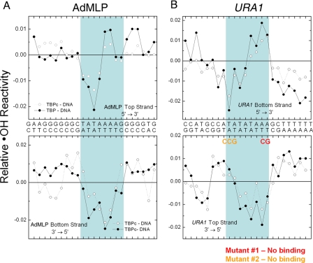
FIGURE 6.
Hydroxyl radical footprinting of the TBP-URA1 DNA complex. The graphs show the extent of hydroxyl radical DNA cleavage of TBPc-DNA (open circles) and TBP-DNA (closed circles) complexes formed on the AdMLP (A) and URA1 (B) DNAs. The blue box in A indicates the AdMLP DNA sequence bound by TBPc in the co-crystal structure (6, 22). The blue box in B shows the DNA sequence contacted by TBP and TBPc in the URA1 promoter inferred by the effects of the TATA mutants and the similarities with the AdMLP footprinting patterns. Note that compared with the AdMLP, the URA1 sequence is inverted with respect to the direction of transcription.

