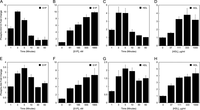
FIGURE 3.
HDL stimulates Erk1/2 and Akt activation in endothelial cells. The effect of S1P and HDL on activation of Erk1/2 (A-D) and Akt (E-H) in HUVEC was determined by multiplex bead array assay. In A-D, the values for the -fold difference in Erk1/2 phosphorylation were derived from the level of phospho-Erk1/2 fluorescence in S1P- or HDL-treated cells divided by the level of phospho-Erk1/2 fluorescence in control cells. In E-G, the values for the -fold increase in Akt phosphorylation were derived from the level of phospho-Akt fluorescence in cells treated with S1P or HDL divided by the level of phospho-Akt fluorescence measured in control cells. The data depicted in panels A, C, E, and G is based on treating HUVEC for the indicated times with 833 nm S1P or 333 μg/ml HDL (containing 133 nm S1P). The data depicted in panels B, D, F, and H are based on treating HUVEC with the indicated concentrations of S1P or HDL for 3 min. The level of S1P in the 3-fold dilutions of HDL tested in panel H ranged from 12 to 337 nm. Data are shown from a representative experiment. Each experiment was performed two times; each data point is an average from two independent wells.

