Figure 2.
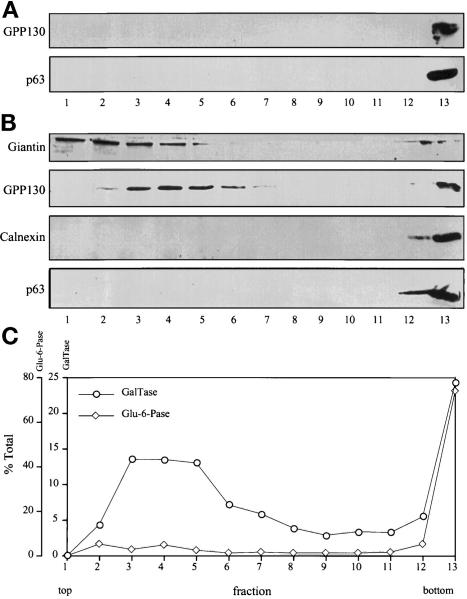
Distribution of ER and Golgi markers after velocity gradient sedimentation. (A) Interphase (nonsynchronized) HeLa cell postnuclear supernatants were fractionated on glycerol gradients and immunoblotted with antibodies against the Golgi marker protein GPP130 and the ER marker protein p63. Both Golgi and ER marker proteins were recovered on a sucrose cushion at the bottom of the gradient. (B) Mitotic HeLa cell postnuclear supernatants were fractionated and immunoblotted with antibodies against Golgi marker proteins (GPP130 and giantin) and ER marker proteins (calnexin and p63). The cells were blocked at mitosis with 0.5 μg/ml nocodazole and collected by shake-off. In contrast to interphase, a major portion of the mitotic Golgi was recovered in a slowly sedimenting position near the top of the gradient. These fractions lacked detectable ER. (C) Fractions resulting from separation of mitotic postnuclear supernatants were also assayed by enzymatic activity for the Golgi marker galactosyltransferase and the ER marker glucose-6-phophatase. By activity assay also, mitotic Golgi, but not mitotic ER, was recovered in a slowly sedimenting position. Centrifugation in these experiments was for 30 min at 150,000 × g; similar results were obtained under a variety of centrifugation conditions.

