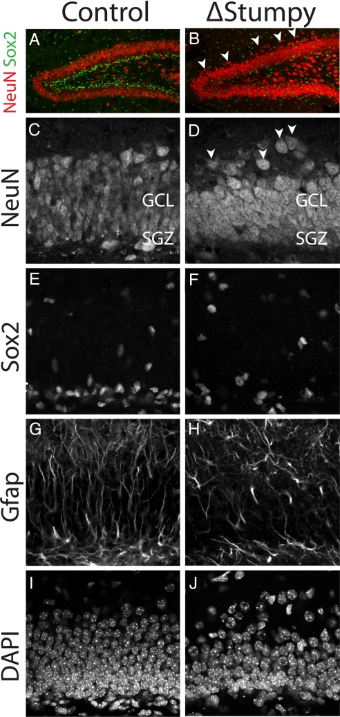
Fig. 1.
Gross hippocampal defects in Stumpy-deficient brain. (A and B) Immunostaining at P13 for NeuN revealed an overall smaller granule cell layer (GCL) and dispersed neurons (white arrowheads) in Stumpy-mutant (ΔStumpy) compared to control brains. (C and D) NeuN-labeled neuronal nuclei showed reduced thickness of the GCL in mutant (D) compared to heterozygous littermates (C). White arrowheads denote dispersed granule cells. (E and F) Sox2-labeled nuclei were dispersed in ΔStumpy (F) compared to control (E). (G and H) GFAP immunostaining in ΔStumpy brain (H) revealed a dramatic reduction of radial glia and fiber density compared to control (G). Remaining GFAP+ cells in ΔStumpy mice had abnormal morphology characterized by disorganized and misoriented processes. (I and J) DAPI-nuclear staining showed decreased cell density in both the SGZ and GCL of mutants (J) compared to control (I).

