Fig. 2.
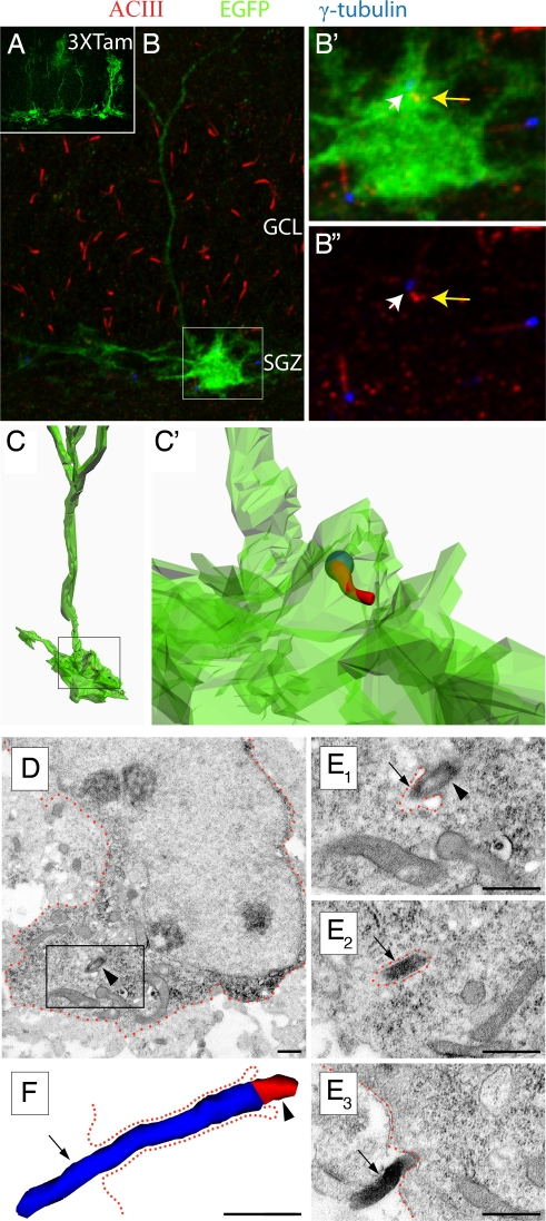
Cilia are present on hippocampal ALNPs. (A) GFAP-CRE-EGFP transgenic mice were injected with tamoxifen for three days and sacrifed one day after the last injection. An example of mosaic labeling of glia with EGFP is shown. (B) EGFP-expressing radial glial cell (boxed) in the subgranular zone (SGZ) in conjunction with immunstaining for ACIII (red) and γ-tubulin (blue). Cilia were located in both GCL and SGZ. (B′) Higher magnification of the inset in B. A cilium was observed extending from the EGFP+ cell expressing both γ-tubulin (white arrow) and ACIII (yellow arrow). (B″) The inset in B showing γ-tubulin (white arrow) and ACIII (yellow arrow). Note that the cilia length is much shorter than overlying GCL cells. (C, C′) Z-stack data were used to reconstruct the cell in B and the location of the cilum within the cell membrane (C′). The spatial resolution of this technique is limited by the technology. Nevertheless, the cilium appears to be largely enveloped in the EGFP+ membrane. (D) An astrocyte glial cell harboring a cilium in the SGZ of a P7 wild-type mouse. Low power electron micrograph of a GFAP+ cell detected with diffuse anti-GFAP DAB immunoprecipitation in the cytoplasm. The framed area is depicted in serial high power micrographs in E. (E) Examples of the basal body (arrowhead) and the ACIII+ axoneme (arrow) detected with black Ni-intensified DAB-immunoprecipitation. (F) Three-dimensional-reconstruction of the cilium showing the basal body (red; arrowhead) and the axoneme (blue; arrow). The cilium was followed in 12 consecutive serial sections until its termination. The total length of the axoneme (∼1.7 μm) was measured in 3D reconstruction. The cell membrane is approximately outlined by green dotted lines. Scale bars, 0.5 μm.

