Figure 3.
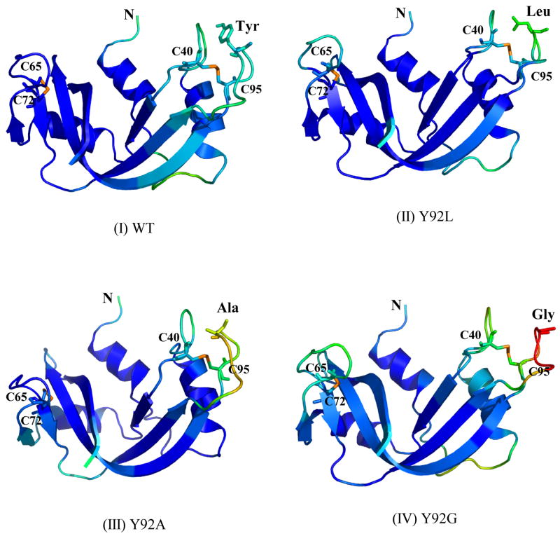
Figure 3A. Crystal structures of (I) WT, (II) Y92L (1YMN), (III) Y92A (1YMR), and (IV) Y92G (1YMW) RNase’s. The residues at position 92, the (65–72) and (40–95) disulfide bonds in WT RNase A and its mutants are depicted using “sticks”. The structures are colored according to the values of the temperature factors (tf) using the equal-sized tf-value increments in the “BGR” (blue, green, and red) color code (10 < tf < 70).
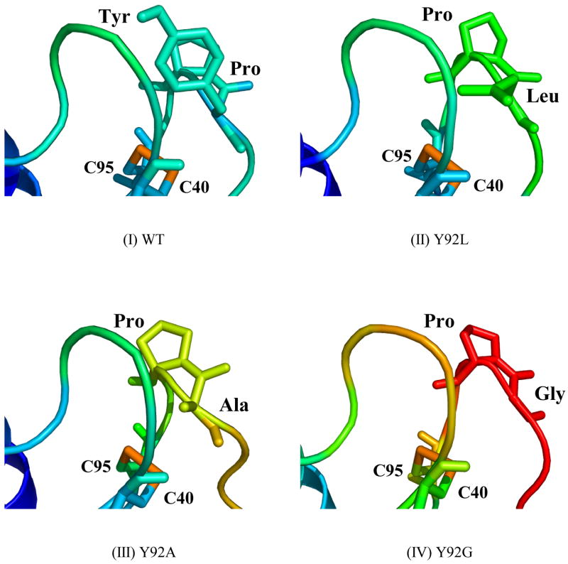
3B. Enlargements of the loop regions containing the (40–95) disulfide bond in (I) WT, (II) Y92L (1YMN), (III) Y92A (1YMR), and (IV) Y92G (1YMW) RNase’s. Pro93 and the residue immediately preceding it are depicted using “sticks”. The structures are colored according to the values of the temperature factors (tf) using the equal-sized tf-value increments in the “BGR” (blue, green, and red) color code (10 < tf < 70).


