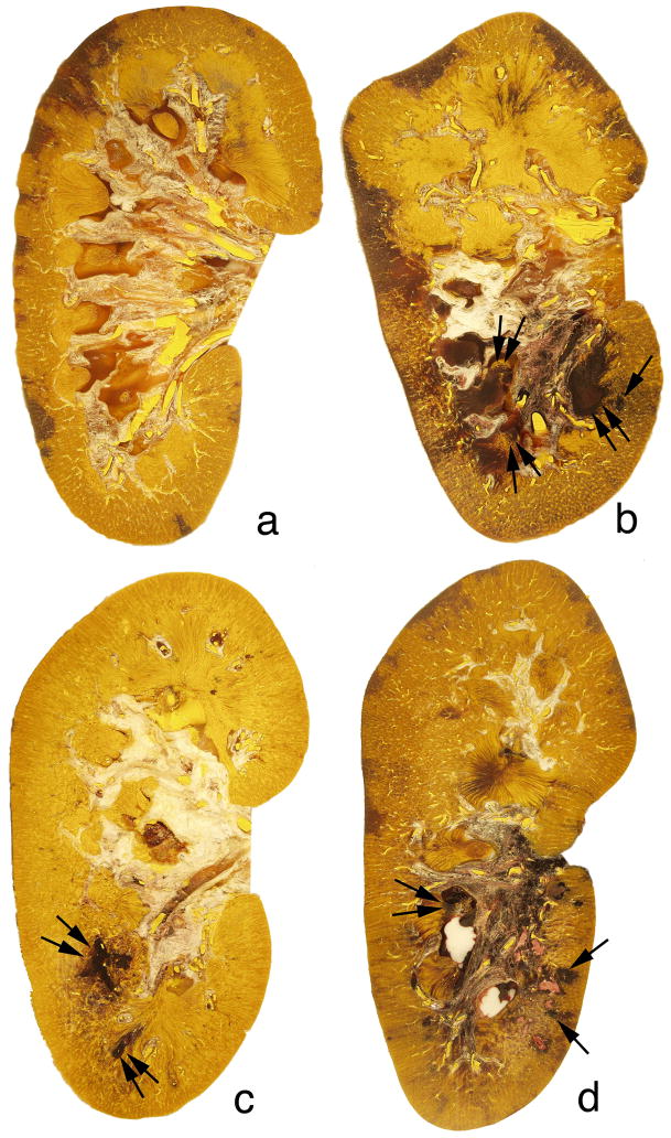
Figure 3.
Digitized and colorized cross-sections of Duet-treated and HM3-treated kidneys. The degree of intraparenchymal hemorrhage induced by the Duet lithotripter varied from no detectable lesion in one animal (panel a) to a lesion size of 3.16% of FRV (panel b). The sites of intraparenchymal hemorrhage were noted in the papilla (double arrows) and adjacent cortical tissue (arrow) within the focal zone. All kidneys that received 2400 SWs (panel c) or 4800 SWs (panel d) from the HM3 lithotripter had lesions similar to that seen in the Duet-treated kidneys. Sites of intraparenchymal hemorrhage were seen in the medulla (double arrow) and cortex (arrows). Magnification, ×1.2 (a–d).

