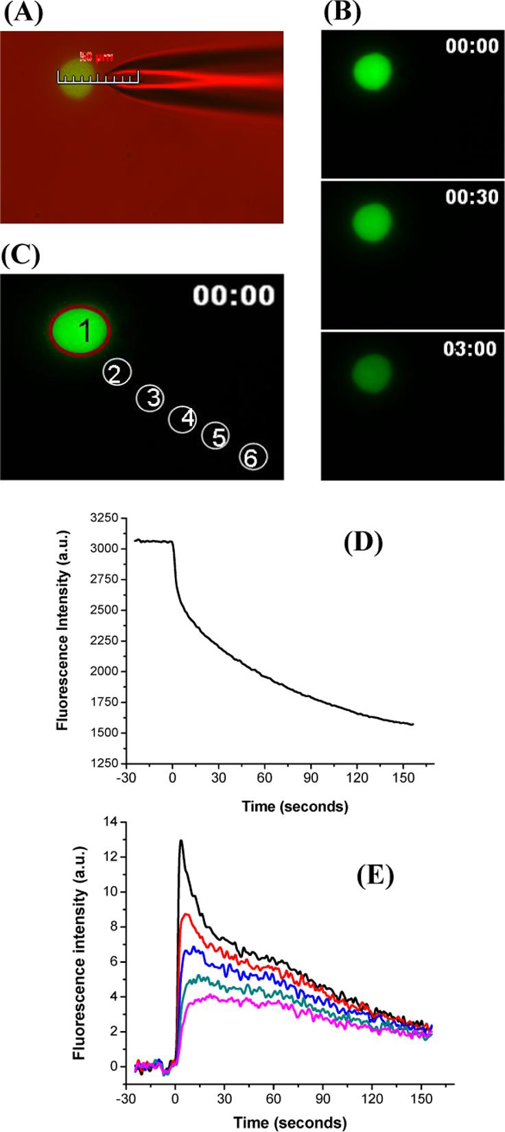
Figure 2.

Visualization of single cell electroporation. Plated cells were incubated with 2 μM Thioglo-1 in the intracellular buffer. A single pulse of 200 ms at a cell-capillary tip distance 5.0 μm was applied at 500 V. Images were collected at a frequency of one frame per second. (A) Photomicrograph produced by an overlay of fluorescence and differential interference contrast image. The image shows the placement of the capillary at a distance of 5 μm from the cell. (B) Fluorescence images before pulsation (0 s), and after pulsation (30 s and 3 min from the start of acquisition). (D) Change in average fluorescence intensity for region 1 (C) against time. (E) Change in average fluorescence intensity for regions 2−6 (C) outside the cell against time. All the data were corrected for bleaching.
