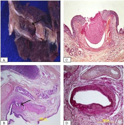
Figure 2.

Pathological Findings. Macroscopic view showing a minute defect within the bronchial tree (arrow) (A). Low-power view showing the location of the vessel in the sub-mucosa beneath the cartilage plate, and the presence of the material of embolization in the lumen (arrow) (B). High-power view revealing the protrusion of the superficial vessel in the lumen with an ulceration and a squamous metaplasia of the epithelium (C). High power-view showing a dysplastic artery with elastic stain (Miller stain) (D).
