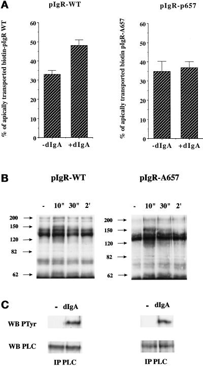
Figure 11.
dIgA binding to pIgR-A657 stimulates PTK activation but not step 3 of transcytosis. (A) Quantitations of the postmicrotubule-dependent assay performed according to the protocol presented in Figure 1 with MDCK cells expressing either pIgR-WT or pIgR-A657. (B) After exposure to dIgA (0.3 mg/ml) for the indicated period, the cells were lysed, and the lysates immunoprecipitated with a specific anti-phosphotyrosine mAb (4G10). The immunoprecipitates were resolved on SDS-PAGE, the gel was transferred to a PVDF membrane, and the membrane was probed with the anti-phosphotyrosine antibody. The arrows indicate bands that exhibit an increase in tyrosine phosphorylation. (C) After a 30-s exposure to dIgA (0.3 mg/ml), the cells were lysed, and the lysates were immunoprecipitated with a mixture of monoclonal anti-PLC-γ1 antibodies. The immunoprecipitates were resolved on SDS-PAGE, the gel was transferred to a PVDF membrane, and the membrane was first probed with the anti-phosphotyrosine antibody (top gels). After stripping, the membrane was reprobed with the same anti-PLC-γ1 mAbs (bottom gels).

