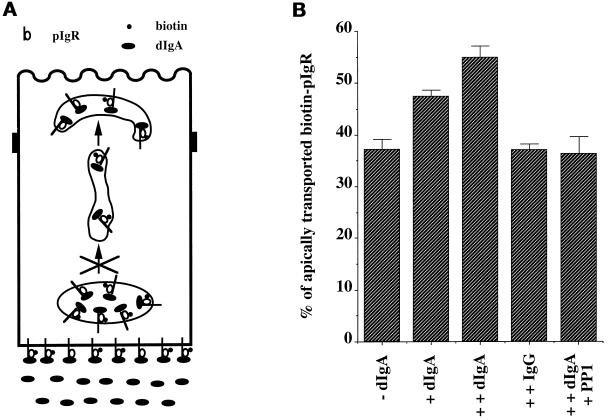
Figure 5.
Basolateral dIgA stimulates apical transport of pIgR. (A) The cells were treated according to the protocol presented in Figure 1 with the modifications shown and described in the text. (B) Quantitation of the apical transport of pIgR in the presence or absence of dIgA added at stage B or in the presence of basolateral dIgA or IgG at stages B and E. n = 5; p < 0.003 for +dIgA; p < 0.001 for ++dIgA; p > 0.5 for ++IgG and ++IgA + PP1.

