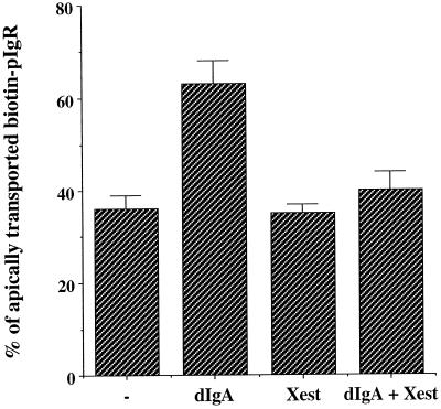
Figure 6.
The increase of intracellular calcium concentration stimulated by dIgA is controlled by the IP3-R. MDCK cells expressing pIgR-WT were submitted to the postmicrotubule-dependent assay as presented in Figure 1. As described in MATERIALS AND METHODS, the assay in this figure as well as Figures 6 and 9 was simplified by omitting the nocodazole incubation (stage D) and combining stages C and E into one incubation at 37°C for 30 min. When indicated the cells were exposed basolaterally to dIgA at stage B. When indicated cells were pretreated with xestospongin C (10 μM) 30 min before biotinylation. The drug was also present throughout the assay. n = 3; p < 0.001 for dIgA; p > 0.5 for Xest and dIgA + Xest.

