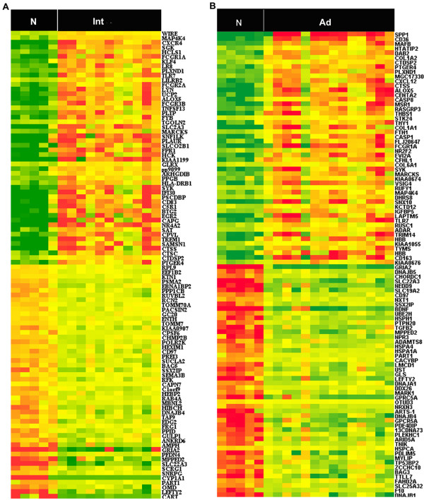
Figure 2.
Heatmap of the 100 most differentially expressed genes in intermediate lesions and advanced lesions, respectively. SAM analysis reveals genes with differential expression in PAD. This analysis compared plaques from within arteries of either intermediate (n = 11) or advanced lesions (n = 14) to normal control group, respectively. Heatmap representation of the 100 most differentially expressed in intermediate lesions (A) and advanced lesions (B). Samples are displayed in columns and genes in rows. Gene expression is represented as a color, normalized across each row, with brighter red for higher values and brighter green for lower values. Gene symbols are listed to the right. N (Normal control group), Int (intermediate lesions group), Ad(advanced lesions group). The list of differentially expressed genes in intermediate lesions and advanced lesions is provided [see Additional file 1 and Additional file 4].

