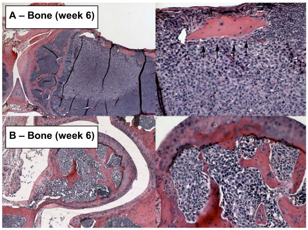
Figure 6.
Metastasis to bone. (Top Left) Metastasis near joint between femur and tibia at week 6. Note extensive degradation of bone adjacent to the upper surface of the tumor. (Top Right) Interface between tumor and bone at higher magnification. Note osteoclasts (arrowheads) lining the lower surface of the bone. (Bottom Left and Right) Femoral metastasis within the joint itself at low and high resolution at 6 weeks. Specimens were obtained from the experiment described in Figure 3.

