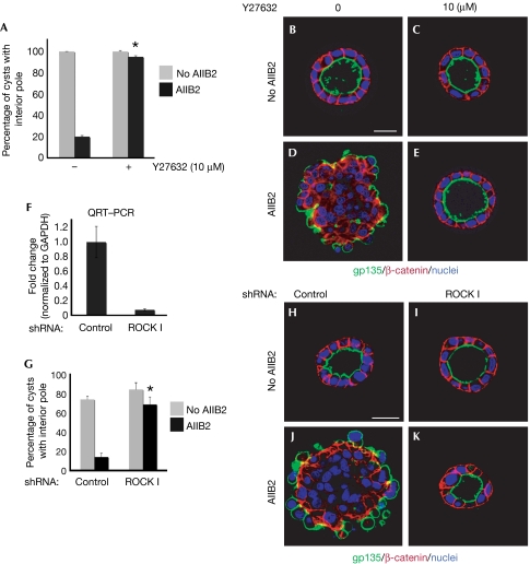
Figure 2.
Depletion of ROCK I rescues AIIB2 polarity. (A) Quantification of cysts with normal polarity in cells treated with Y27632; *P<0.001. (B–E) Representative confocal images of cysts stained with gp135 (green) and β-catenin (red); nuclei are shown in blue. (F–K) Depletion of ROCK I rescues AIIB2-induced phenotype. (F) Reduction of ROCK I expression level by RNAi is confirmed by QRT–PCR. (G) Quantification of cysts with normal polarity in cells transfected with shRNA to ROCK I; n=3; *P<0.001. (H–J) Confocal images of representative cysts. Cells infected with lentivirus for control or ROCK I were plated in COLI for 5 days and stained with gp135 (green) and β-catenin (red); nuclei are shown in blue. Scale bars, 20 μm. COLI, collagen I; QRT–PCR, quantitative reverse transcription–PCR; RNAi, RNA interference; ROCK, Rho kinase; shRNA, short hairpin RNA.

