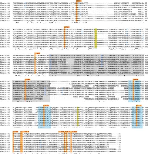
Figure 2.
Conserved tyrosine residues in the cytoplasmic domain of plexin family members. Alignment of protein sequences of human plexin intracellular domains. In the consensus line (shown under the sequences), amino-acid identity is marked with asterisks and similarity is indicated by dots or columns, as assigned by the CLUSTALW algorithm. Background colours highlight residues or domains of particular interest. In total, 13 tyrosine residues (Y) are conserved in all plexins (or in all but one family member) and are shown in red; moreover, three of these residues are included in highly conserved amino-acid stretches (blue background). The positions of highly conserved tyrosines are indicated on top of the sequences, with reference to the amino-acid coordinates in plexin A1 (accession number NP-115618.2). Notably, the residues diverging from this consensus in plexin B3 and plexin A4 (underlined) are conserved between the human and mouse genes. Tyrosine residues that are conserved in at least one entire plexin subfamily are shown in blue. The three conserved arginines found to be functionally required in the GTPase-activating protein-like motifs are shown in green (Vikis et al, 2002). A grey background highlights the presumptive RhoGTPase-binding domain (Tong et al, 2008).

