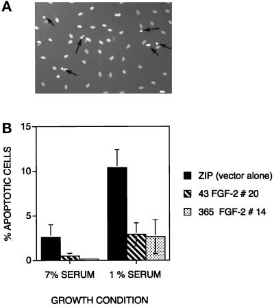
Figure 8.
Apoptosis in cells from clones expressing LMW or HMW FGF-2 forms. (A) Representative Hoechst 33342 staining of LMW FGF-2–expressing cells maintained 4 d in 1% serum. Apoptotic nuclei are brighter and smaller than normal nuclei as a result of chromatin condensation (indicated by arrows). Original magnification, 200×. (B) Histogram presenting percentage apoptotic cells in cultures incubated in complete and serum-depleted medium. Three fields of at least 300 cells each were counted for each sample. Data represent the mean ± SD from triplicate samples.

