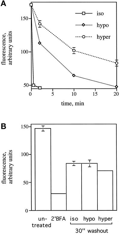
Figure 9.
Effect of hypotonic and hypertonic treatment on the rate of βCOP dissociation (A) and association (B). (A) HeLa cells, grown on 12-mm glass coverslips, were left in normal medium without serum or placed in hypotonic medium (210 mOsm) or hypertonic medium (610 mOsm) for 1 min before the addition of 10 μg/ml BFA. At each time point indicated, cells were fixed and stained using a mAb against βCOP and a polyclonal antiserum against giantin. The average of the mean fluorescence intensity of βCOP staining in a fixed circle (2782 pixels) enclosing the Golgi region (as indicated by giantin staining) in 50 cells was measured. (B) HeLa cells on coverslips were incubated with 2.5 μg/ml BFA in HEPES-buffered normal medium, pH 7.2, without serum for 2 min at room temperature to induce the dissociation of βCOP from the Golgi. Rebinding of βCOP to the Golgi was assessed by washing cells four times with 1 ml of normal medium without serum, hypotonic medium (210 mOsm), or hypertonic medium (610 mOsm) and placing them at 37°C for 30 s. The average of the mean fluorescence intensity of βCOP staining was determined as in A. Vertical bars represent the SEM as determined from 25 measurements.

