Figure 2.
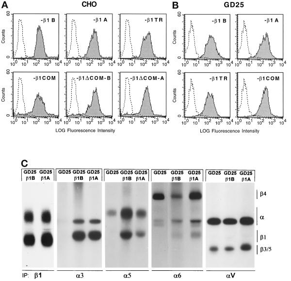
Surface expression of transfected β1 variants in CHO and GD25 cells. CHO and GD25 cells were transfected with DNA constructs for human β1B or β1A or for the β1 cytoplasmic domain deletion mutants indicated, and positive cells were sorted by panning on β1 antibodies as described in MATERIALS AND METHODS. (A and B) To detect surface expression of transfected β1 variants, CHO (A) and GD25 (B) transfectants were stained with mAb TS2/16 to human β1, followed by fluorescein-labeled anti-mouse IgG, and analyzed by FACS. The level of β1 surface expression reported as fluorescence intensity is shown. Untransfected cells were included as a negative control. (C) Integrin heterodimers in 125I-surface-labeled GD25, GD25-β1B, and GD25-β1A cells as detected by nonreducing SDS-PAGE and autoradiography of immunoprecipitated integrins with antibodies specific for β1, α3, α5, α6, and αV subunits.

