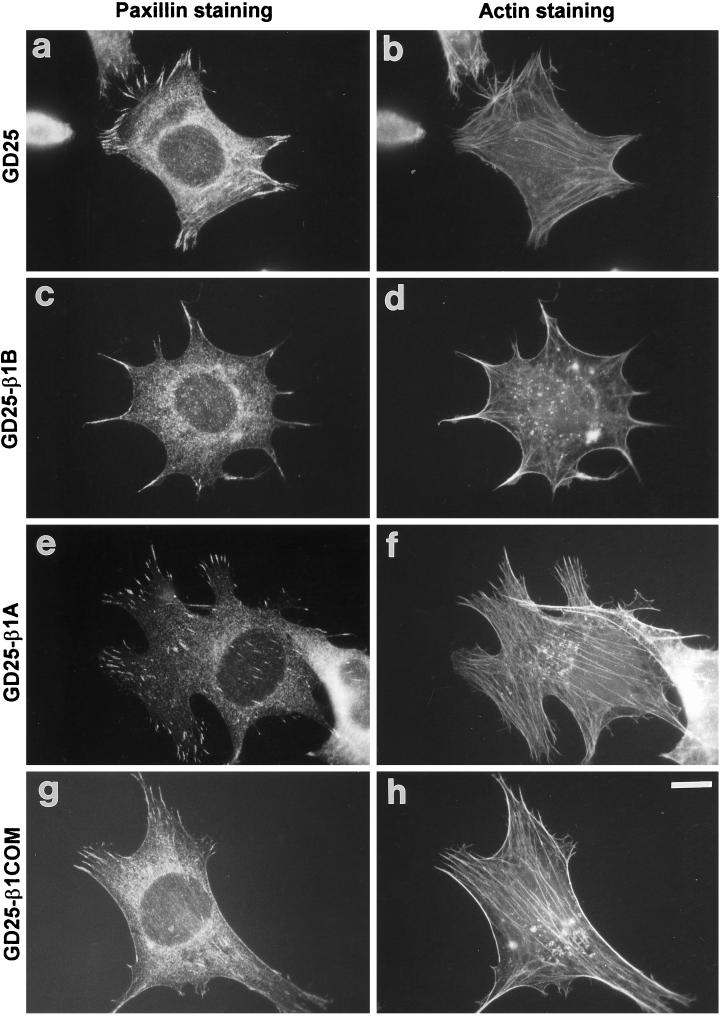
Figure 8.
Focal adhesion and stress fiber organization in control and transfected GD25 cells. Cells were plated on 10 μg/ml fibronectin-coated glass coverslips for 3 h in complete culture medium. Cells were then fixed for 10 min in 3.7% (v/v) paraformaldehyde in PBS, permeabilized with 0.5% Triton X-100 and 3.7% formaldehyde in PBS for 5 min, and double stained for paxillin (A, C, E, G) and F-actin (B, D, F, H) as described in MATERIALS AND METHODS. Note the reduction of stress fibers and paxillin-containing focal adhesions in GD25-β1B cells. Bar, 10 μm.

