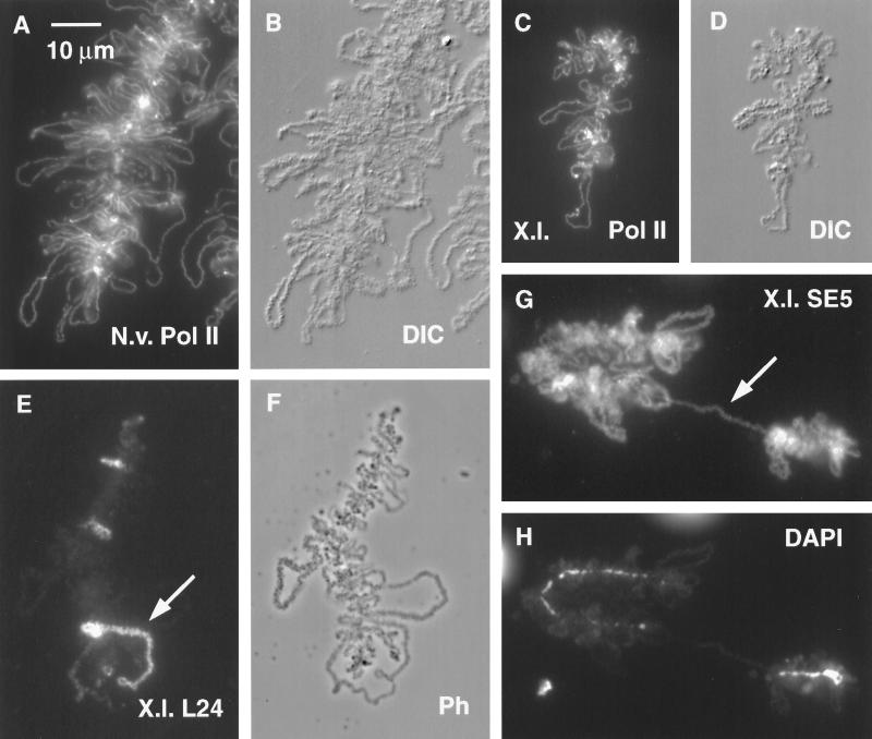
Figure 8.
(A) Small portion of a typical LBC from a GV of the newt N. viridescens, stained with mAb H14 against RNA Pol II. (B) Same field by DIC. Compared with a Xenopus LBC, the newt LBC has longer loops, more heavily textured loop matrix, and unusually distinct Pol II (+) loop axes. In addition, newt LBCs are longer than Xenopus LBCs. (C) LBC derived from a Xenopus sperm head that had been injected 2 d previously into a newt GV. Sample was stained for Pol II. (D) Same field by DIC. Note how strongly this Xenopus LBC resembles a newt LBC except for length. (E) Xenopus sperm LBC 2 d after injection of Xenopus sperm into a newt GV. Sample was stained with serum L24 against xnf7 (Reddy et al., 1991). Only a few loops react strongly with this antibody. The large loop (arrow) near the chromosome end is single, not paired like the loops of an oocyte LBC. This loop consists of two tandem transcription units, only one of which stains. (F) Same field by phase contrast. (G) Xenopus sperm LBC 2 d after injection into a newt GV. Sample was stained with mAb SE5 (Roth and Gall, 1987), which normally stains newt but not Xenopus LBC loops. Staining demonstrates that newt proteins were used to assemble the Xenopus LBC. Arrow indicates a single-loop bridge described in the text. (H) Same field (DAPI stain) showing that the loop bridge spans a gap in the chromomere axis.

