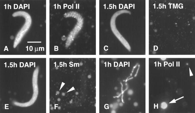
Figure 9.
(A) Xenopus sperm head 1 h after injection into a Xenopus GV. The oocyte was incubated in actinomycin D (20 μg/ml) for 1 h before injection and was returned to the drug after injection (DAPI stain). (B) Same sperm head stained with mAb H14, showing uptake of Pol II by the sperm even though transcription was inhibited. (C) Sperm head 1.5 h after injection into an actinomycin-treated oocyte (DAPI stain). (D) Same sperm head stained with mAb K121 against TMG, showing the absence of splicing snRNAs. (E) Sperm head 1.5 h after injection into an actinomycin-treated oocyte (DAPI stain). (F) Same sperm head stained with mAb Y12, showing absence of Sm proteins. Arrowheads point to Y12 (+) B-snurposomes. (G) Highly contracted chromosome from the same GV that contained the sperm head shown in A and B (DAPI stain). (H) Same area showing absence of chromosomal stain with mAb H14, except for the terminal granule (arrowhead). A coiled body (sphere) shows typical staining (arrow).

