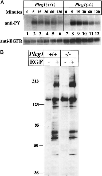
Figure 2.
Influence of PLC-γ1 on EGF receptor autophosphorylation and protein tyrosine phosphorylation. (A) Confluent Plcg1+/+ and Plcg1−/− cells were incubated overnight in DMEM plus 0.5% fetal bovine serum and then treated at 37°C with EGF (20 ng/ml) for the indicated times. Cells were then lysed, and the EGF receptor was immunoprecipitated and probed by Western blotting with antiphosphotyrosine as described in MATERIALS AND METHODS. (B) Confluent Plcg1+/+ and Plcg1−/− cells were incubated in DMEM plus 0.5% fetal calf serum overnight, and the cells were then treated for 5 min at 37° without or with EGF (50 ng/ml). After cell lysis, equal aliquots (40 μg) of each lysate were subjected to SDS-PAGE and Western blotting with antiphosphotyrosine. Bound antibody was detected by ECL.

