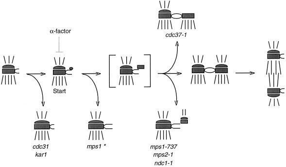
Figure 9.
Proposed pathway for SPB duplication. In this schematic, the central and outer plaques of the SPB are depicted, with cytoplasmic and nuclear microtubules drawn above and below the SPB, respectively. The bracketed structure is a duplication intermediate that is inferred but has not been reported. Execution point experiments with mps1–1, mps2–1, and ndc1–1 mutants (Winey et al., 1991, 1993) indicate where in the pathway these mutations cause SPB duplication to fail. The mps1–1 mutation causes failure early on and is marked with an asterisk to indicate that four of the other five alleles share this SPB morphology. However, the mps1–737 mutation allows duplication to proceed farther before failure occurs and has been placed here at the same point in the pathway as mps2–1 and ndc1–1 because of their phenotypic similarity. Mutation of CDC37 also causes a later failure in SPB duplication, but generates a different, partially duplicated structure (Schutz et al., 1997). The two distinct mps1 mutant phenotypes suggest that this gene is required continuously or at multiple times during SPB duplication.

