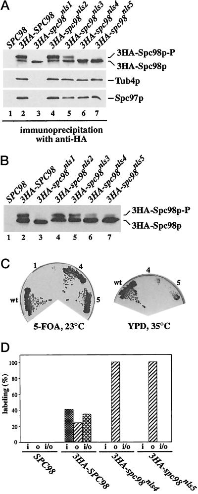
Figure 6.
The cytoplasmic Spc98pnls4 and Spc98pnls5 are not hyperphosphorylated and associate with the outer plaque of the SPB. (A) 3HA-Spc98pnls4 and 3HA-Spc98pnls5 form complexes with Tub4p and Spc97p. Proteins of cells expressing SPC98 only (lane 1), or SPC98 together with either 3HA-SPC98 (pSM338), 3HA-spc98nls1 (pGP20), 3HA-spc98nls2 (pGP22), 3HA-spc98nls3 (pGP27), 3HA-spc98nls4 (pSM456), or 3HA-spc98nls5 (pGP31) (lanes 2–7) were precipitated using anti-HA antibodies. Spc98p, Spc97p, and Tub4p were detected in the precipitate by immunoblotting using affinity-purified polyclonal anti-Spc98p, anti-Tub4p, or anti-Spc97p antibodies. (B) Spc98pnls4 and Spc98pnls5 can become phosphorylated. Extracts of cells of ESM243 (Δspc98::HIS3 pRS316-SPC98) carrying plasmid pRS425 (lane 1), pGP43 (3HA-SPC98), pGP44 (3HA-spc98nls1), pGP45 (3HA-spc98nls2), pGP46 (3HA-spc98nls3), pGP58 (3HA-spc98nls4), or pGP47 (3HA-spc98nls5) (lanes 2–7) were prepared, followed by immunoprecipitation using anti-HA antibodies. The precipitates were analyzed by immunoblotting using polyclonal anti-Spc98p antibodies. These cells carried in addition SPC97 and TUB4 on the 2-μm plasmid pGP48. (C) Spc98pnls4 and Spc98pnls5 become functional after overproduction. Cells of strain ESM243 with plasmids pGP48 and either pGP43 (3HA-SPC98; sector wt), or pGP44 (3HA-spc98nls1, sector 1), or pGP58 (3HA-spc98nls4, sector 4) or pGP47 (3HA-spc98nls5, sector 5) were grown on 5-FOA at 23°C. 5-FOA selects against the cells carrying the plasmid pRS316-SPC98. Growth on this plate indicates that the SPC98 derivative is functional. Cells from the 5-FOA plate were streaked out on a YPD plate. Cells with 3HA-SPC98 (sector wt) grew, while 3HA-spc98nls4 (sector 4) and 3HA-SPC98nls5 (sector 5) cells failed to grow at 35°C, but grew at 23°C (our unpublished result). (D) 3HA-Spc98pnls4 and 3HA-Spc98pnls5 are associated with the outer plaque of the SPB. Cells expressing SPC98 or SPC98 together with 3HA-SPC98, 3HA-spc98nls4, or 3HA-spc98nls5 were prepared for immunoelectron microscopy using anti-HA antibodies as described (Knop et al., 1997). The distribution of gold particles associated with 25 SPBs was determined in each case. The HA signal was associated with the inner (i), outer (o), or inner and outer (i/o) plaques but not with other SPB substructures.

