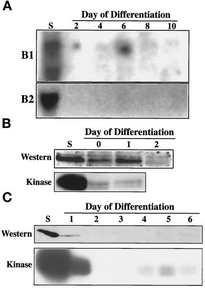
Figure 6.
Expression of the B-type cyclins during differentiation of the Rcho-1 cells. (A) Total RNA was prepared from the Rcho-1 cells at the times indicated, and analyzed for cyclin B1 and B2 expression by hybridization to the full-length probes as indicated. (B) Analysis of expression and activity of the cyclin B protein products during the first endocycle. Lysates were prepared in an RIPA buffer from proliferating stem (S) or differentiating giant cells on the d indicated in the panel. Equivalent amounts of the proteins were separated on a 10% polyacrylamide gel, transferred, and the proteins were detected by the electrochemiluminescence system using the anti-cyclin B antibody. The lysates shown in the two panels were derived from two different experiments. Matching samples of the lysates used in the analysis of cyclin B protein expression were subjected to immunoprecipitation using the same anti-cyclin B1 antibody, and the immunoprecipitates were assayed for histone H1 kinase activity. The products were separated on a 12.5% polyacrylamide gel and the autoradiograph is shown. (C) Analysis of cyclin B protein expression and associated kinase over an extended time course. Lysates were prepared and analyzed for cyclin B abundance and associated kinase activity, as described in B from the samples indicated.

