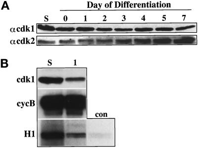
Figure 7.
Expression of cdk1 and cdk2 and their association with cyclin B1 during the endocycle. (A) Analysis of of p34cdk1 and p33cdk2 expression during Rcho-1 differentiation. Protein lysates derived from the differentiating cells were separated and analyzed by immunoblotting for the presence of the two cdks in proliferating (S) and differentiating cells at the time points indicated. The membranes were probed with the antibody indicated to the left of each panel (αcdk1 = anti-p34cdk1 antibody; αcdk2 = anti-p33cdk2 antibody) and the presence of the protein was revealed by electrochemiluminescence (Amersham). (B) Reduction of p34cdk1 and cyclin B1 association with the onset of differentiation. Immunoprecipitates were prepared from lysates of proliferating or differentiating cells on d 1 using the anti-cyclin B1 antibody, in what was estimated to be antigen excess based on previous analyses, and the immunoprecipitates were subjected to either an immunokinase assay (H1) or an immunoblot to determine the amount of cyclin B1 (B) or associated p34cdk1. The positions of the cyclin B1 and p34cdk1 are indicated on the left of the panels.

