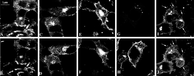
Figure 2.
Subcellular distribution of MMIα in BWTG3 cells by confocal immunofluorescence analysis. Paraformaldehyde-fixed and saponin-permeabilized cells were double labeled with phalloidin (A) and anti-Myr 1 antibodies (Tu 30; B). Cells were incubated with biotinylated transferrin 20 min before fixation, permeabilized with saponin, and double labeled with Texas Red-streptavidin (C) and anti-Myr 1 antibodies (Tu 22; D). Note that two antibodies directed against two different domains of Myr 1 decorated similarly punctate structures in the perinuclear region. After preservation of the endocytic compartments according to the procedure of Stoorvogel et al. (1996) (see MATERIALS AND METHODS), cells were paraformaldehyde fixed, detergent permeabilized, and double labeled with anti-Myr 1 antibodies (Tu 30; F, H, and J) and antibody directed against the transferrin receptor (H68.4; E), Golgi complex (G), and β actin (I). Horizontal optical sections throughout the focal plane of the nucleus were obtained by confocal laser scanning microscopy.

