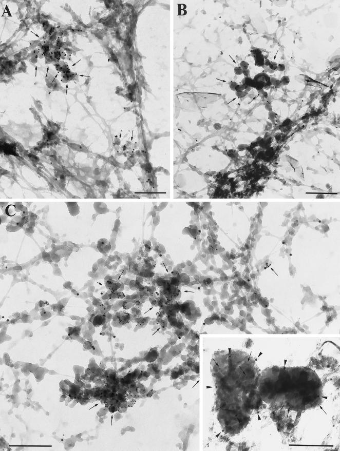
Figure 4.
Visualization of MMIα, the transferrin receptor and lgp 120 by whole-mount EM. (A) DAB-positive tubulo-vesicular structures were intensely labeled with the anti-transferrin receptor antibody (H68.4) directed against its cytoplasmic tail (PAG 10; arrows). (B) Similar DAB-positive tubulo-vesicular structures (endosomes) were labeled with the anti-Myr 1 antibodies (PAG 10, Tu 30; B, arrows). (C) Double immunogold labeling with the anti-Myr 1 antibodies (PAG 10) and the anti-transferrin receptor antibody (PAG 5) showed the codistribution of MMIα and transferrin receptor in the same tubulo-vesicular structures (arrows). (C) Inset, Lgp 120 (PAG 5) was detected in DAB-positive lysosomal compartments (arrows), which were also labeled with the anti-Myr 1 antibodies (PAG 10) (arrowheads). Bars, 200 nm.

