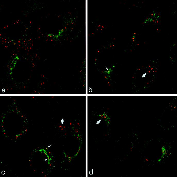
Figure 1.
Double label of M6PR and EGF en route to the lysosome. Cells were incubated with EGF-TxR (red) for 10–60 min at 37°C and were then immunolabeled for M6PR (green). After 10 min at 37°C, EGF-TxR is in vacuoles distributed throughout the peripheral cytoplasm (A). With further incubation at 37°C for 25 (B), 45 (C), and 60 min (D), EGF-TxR accumulates in the juxtanuclear region of the cell (large arrows). At all time points most of the elements labeled with EGF and M6PR are discrete and separate. Small arrows indicate the rare occasions of coincidence between EGF-TxR and M6PR.

