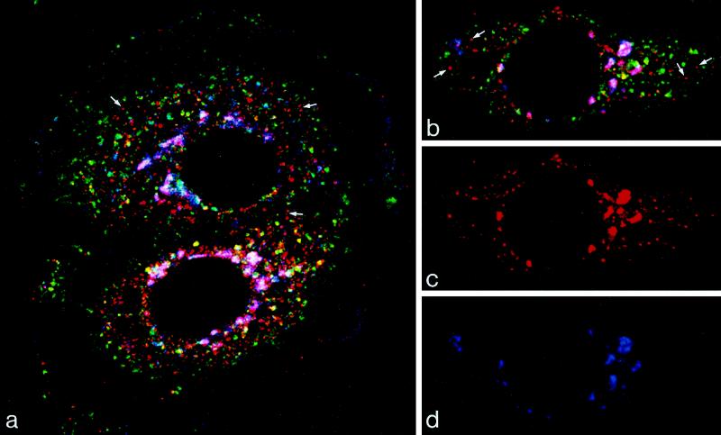
Figure 2.
Triple label showing M6PR in relation to markers of the trans-Golgi and endosome. Cells transiently transfected with ST-HRP were incubated with TF-FITC (green) for 60 min at 37°C and were then immunolabeled for M6PR (red) and HRP (blue). Merged images are shown in A and B, and single images of the cell shown in B are shown in C (M6PR) and D (ST-HRP). M6PR is found in the trans-Golgi region, in which trans-Golgi elements that also contain ST-HRP are stained magenta, and in vacuoles in the peripheral cytoplasm. The majority of the peripheral M6PR-positive vacuoles stain red (arrows) and so do not contain either ST-HRP or TF-FITC. Only a few M6PR-positive vacuoles contain TF (stained yellow), and TF-FITC is not detectable in the trans-Golgi.

