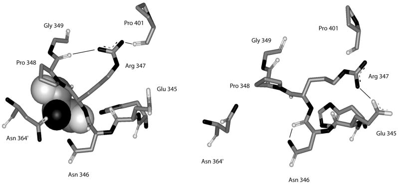
Figure 7. Depiction of the structure of the residues forming the serine binding site of E. coli D-3-Phosphoglycerate Dehydrogenase.
Residues shown are from His-344 to Gly-349 from one subunit and residue Asn-364′ from the adjacent subunit. Hydrogen bonds are shown as solid lines. The structure on the left is from the enzyme in the presence of L-serine and the structure on the right is the enzyme in the absence of L-serine. L-serine is depicted as a CPK structure.

