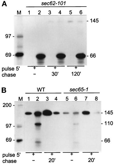
Figure 1.
Secretion of Hsp150Δ-β-lactamase in translocation-defective mutants. Sec62–101 (A, lanes 1–6), WT (B, lanes 1–4), and sec65–1 (B, lanes 5–8) cells (5 × 10⁸/ml) were preincubated for 15 min and labeled with [35S]methionine/cysteine for 5 min. Parallel similarly pulse-labeled cells received CHX and were chased for the indicated times. In A all incubations were performed at 30°C and in B at 37°C. The growth media (lanes with uneven numbers) were separated from the cells (lanes with even numbers), which were lysed. All samples were subjected to immunoprecipitation with anti-β-lactamase antiserum followed by SDS-PAGE analysis. The Hsp150Δ-β-lactamase proteins (kDa) are indicated on the right, and the molecular weight markers (lane M; kDa) on the left.

