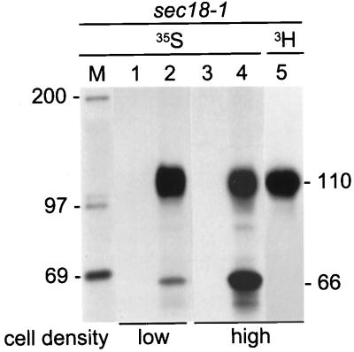
Figure 2.
Retardation of translocation of Hsp150Δ-β-lactamase. (A) Sec18–1 cells were preincubated for 15 min at 37°C and labeled for 1 h at the same temperature with [35S]methionine/cysteine under low cell density conditions (5 × 10⁷/ml; lanes 1 and 2) or high cell density conditions (5 × 10⁸/ml; lanes 3 and 4). One sample was labeled with [3H]mannose under high cell density conditions (lane 5). The media (lanes 1 and 3) were separated from the cells, which were lysed (lanes 2, 4, and 5). The samples were immunoprecipitated with anti-β-lactamase antiserum and analyzed by SDS-PAGE. The Hsp150Δ-β-lactamase proteins (kDa) are indicated on the right and the molecular weight markers (lane M; kDa) on the left. Lanes 3 and 4 were exposed four times longer than were lanes 1 and 2.

