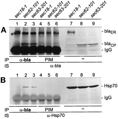
Figure 8.
Coimmunoprecipitation of Hsp70 with cytoplasmic Hsp150Δ-β-lactamase. The indicated yeast strains were incubated under low cell density conditions for 1 h at 37°C (sec18–1) or 30°C (sec62–101 and sec63–201). The sec18–1 cells received CHX and were incubated for an additional 30 min 37°C. NaN3 was added and the cells were lysed under mild detergent conditions. After addition of apyrase, the lysates were immunoprecipitated (IP) with anti-β-lactamase antiserum (α-bla; lanes 1–3, A and B), or with preimmune serum (PIM; lanes 4–6, A and B). The precipitates, and parallel unprecipitated lysate samples (lanes 7–9, A and B), were subjected to SDS-PAGE, followed by Western blotting and immunostaining (IS) with either anti-β-lactamase antiserum (A) or anti-Hsp70 antiserum (B). On the right, ER-located (blaER; 110 kDa) and cytoplasmic (blaCP; 66 kDa) Hsp150Δ-β-lactamase forms, and IgG bands, are indicated. An unspecific band is indicated with an asterisk (A).

