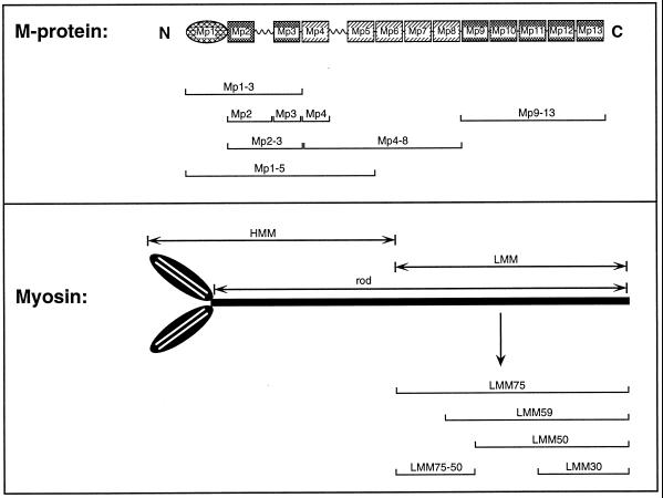
Figure 1.
Summary diagram giving a schematic representation of the domain organizations for M-protein and for myosin. The presentation emphasizes the modular construction of M-protein from repetitive Ig cII (cross-hatched rectangles) and fibronectin type III repeats (shaded rectangles) interspersed by unique sequence stretches of varying length. Recombinant constructs used for mapping of binding sites, transfection of cultured myoblasts, and phosphorylation assays are marked by brackets. The location of the most prominent proteolytic fragments of myosin is given in the lower panel above the myosin sketch. Brackets indicate the borders of the recombinant LMM constructs used in this study.

