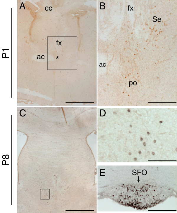
Figure 5.
Pax2 protein expression in the mouse forebrain during early postnatal development. A-E: Coronal forebrain sections immunostained for Pax2 at P1 (A-B) and at P8 (C-E), showing the presence of few disperse Pax2-positive cells in the septal area (Se) (A-C, high power in D), the medial preoptic nucleus (po) (asterisk in A and B), and the subfornical organ (SFO) (arrow in E). The fibre tracts are indicated in A and B for orientation purposes (ac, anterior commissure; cc, corpus callosum; fx, fornix). The boxed areas in A and C delineate the high power images depicted in B and D respectively. Scale bars: A, C, 100 μm; B, E, 25 μm; D, 12.5 μm.

